On the twenty-fifth of April, 1953, the journal Nature carried three short papers on the structure of a single molecule. The first, two pages from two men at Cambridge, proposed a shape so simple it seemed almost a conjuring trick, and closed on the most studied understatement in the history of science. The two papers that followed it came from King's College London and supplied the evidence — and one of their authors had not been told that her photograph, and her unpublished numbers, had already travelled the sixty miles to Cambridge and settled the matter.
The understatement
On a single page, James Watson and Francis Crick set out “a structure for the salt of deoxyribose nucleic acid,” observed that it had “novel features which are of considerable biological interest,” and ended with a sentence that has been quoted ever since: “It has not escaped our notice that the specific pairing we have postulated immediately suggests a possible copying mechanism for the genetic material.”1 A few weeks earlier, by Watson's telling, Crick had walked into the Eagle, the public house across from their laboratory, and announced to the lunchtime room that the pair of them had found the secret of life.2 The story has mostly been told in that key: two young men, an inspired fortnight of cardboard and wire, and the riddle of heredity solved between a Monday and a Friday.
What the famous page does not say plainly, it says once, in a buried line of thanks. The authors had “been stimulated by a knowledge of the general nature of the unpublished experimental results and ideas of Dr. M. H. F. Wilkins, Dr. R. E. Franklin and their co-workers at King's College, London.”3 The word carrying the most weight there is unpublished. The results were not only unpublished. One of the two people named had not been told that the Cambridge pair had seen them.
The shape of the thing
The structure they proposed was two chains wound about a common axis and running, as the paper carefully put it, in opposite directions: a right-handed double helix whose sugar–phosphate backbones lie on the outside and whose bases turn inward, stacked like the rungs of a twisted ladder a little over three and a half ångström apart.4 The rungs are the heart of it. Each is a pair of bases, one drawn from each chain, and only two pairings will fit the geometry — adenine with thymine, guanine with cytosine, a purine always answered by its pyrimidine.5 Erwin Chargaff had reported, without quite knowing what to make of it, that in any sample of DNA the quantity of adenine matched that of thymine, and guanine that of cytosine, almost exactly.6 The double helix accounted for the coincidence at a stroke: if every A is bound to a T and every G to a C, the totals must agree.
And it did more than tidy a ratio. Because each base specifies its partner, the order along one chain dictates the order along the other; part the two strands, and each carries the full instructions for rebuilding its lost half. That is what the closing sentence meant, and why it could afford to be coy. The molecule that carries inheritance had been found to contain, folded into its very geometry, the means of being copied.
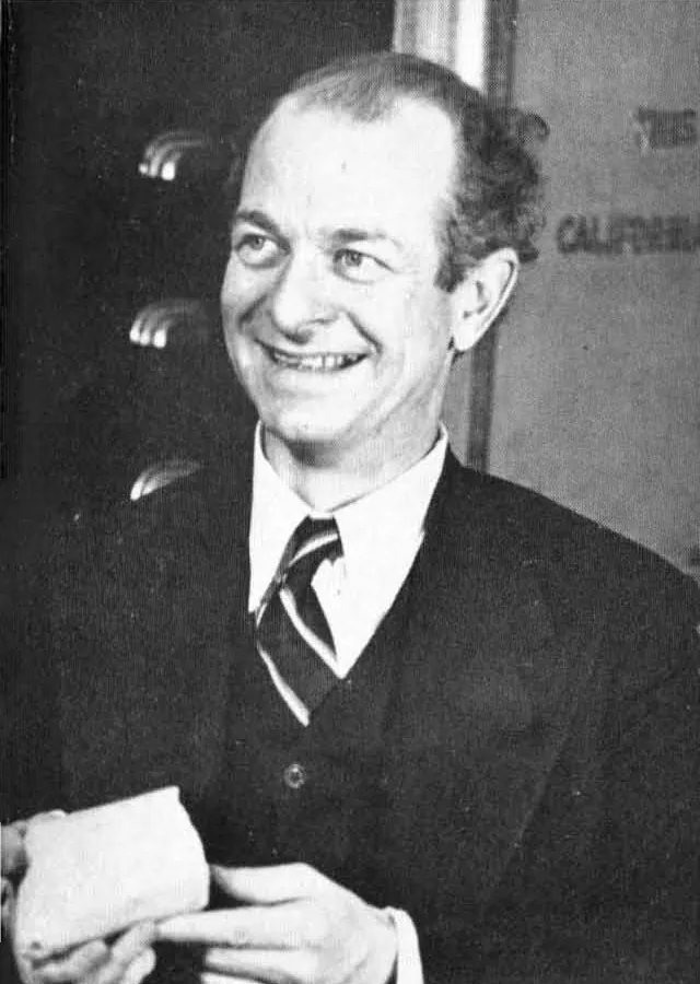
It was not the only structure on offer. Weeks before, Linus Pauling, the foremost chemist alive, had published a model of his own: three chains, not two, the phosphates crowded along the axis.7 It was wrong in a way that should not have been possible for so great a mind. To bind the negative charges he had heaped at the core, Pauling had quietly added hydrogen atoms that, at the acidity of a living cell, are simply not there; he had proposed a form of DNA that was not even an acid. Reading the manuscript Pauling's own son had carried across to Cambridge, Watson saw the blunder, and knew the race was still open.
The fifty-first photograph
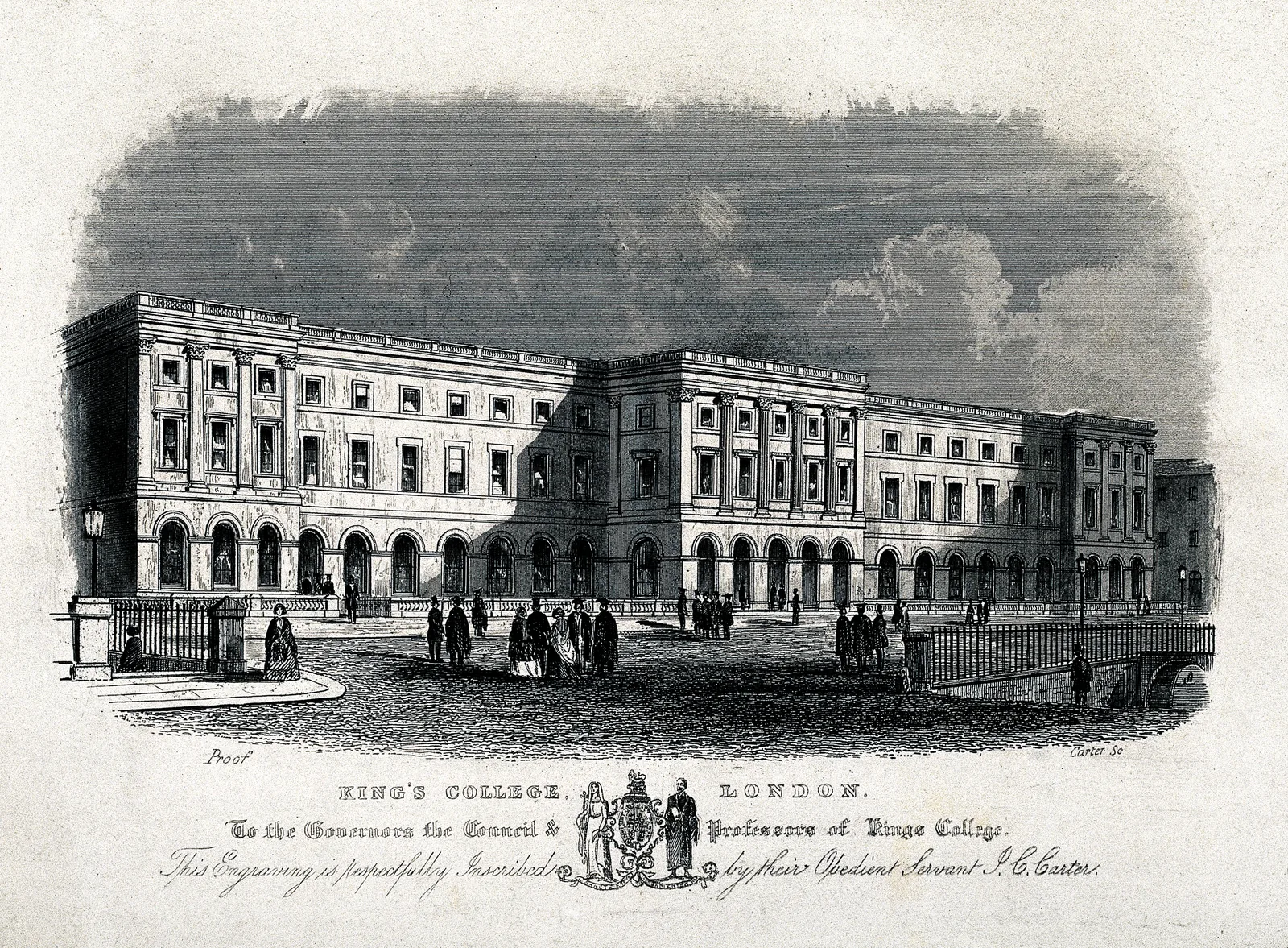
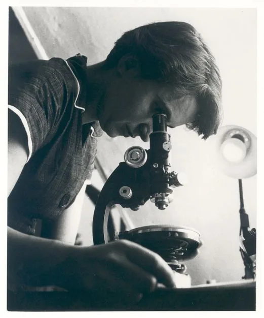
What told against Pauling, and for the double helix, was a body of measurement made far from Pasadena and the Cavendish, in a basement laboratory at King's College London, by a physical chemist named Rosalind Franklin. She had learned the X-ray study of carbons in Paris and become a master of the method, and was brought to King's in 1951 to turn it on DNA.8 With her student Raymond Gosling she rebuilt the apparatus, drew finer fibres than anyone had managed, and held them at controlled humidity — and in doing so established that DNA takes two forms: a short, crystalline “A” form when dry, and a long, extended “B” form when wet.9 In May 1952 she and Gosling trained the X-ray beam on a B-form fibre through a long exposure, and recovered an image whose meaning is legible even to a layman: a broad dark cross of reflections, the unmistakable signature of a helix. It was the fifty-first photograph in their series, and it has been known ever since as Photograph 51.10
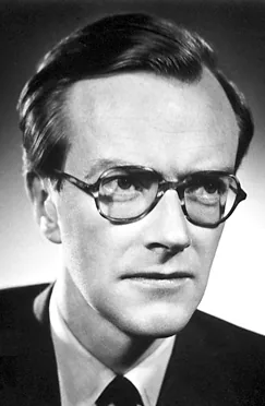
Franklin set it aside. The wet B form was eloquent, but it was the crystalline A form, harder to read and far richer in detail, that she judged would yield a structure that could actually be proved, and she turned to the heavier labour of solving it.11 She was, if anything, too rigorous to leap. And while she worked toward her proof, the photograph she had filed went travelling. In late January 1953 Maurice Wilkins, her nominal colleague (they shared the molecule and almost nothing else), showed Photograph 51 to James Watson, down from Cambridge for the day.12 Watson needed none of the mathematics. “The instant I saw the picture my mouth fell open and my pulse began to race,” he wrote; the cross told him the thing was a helix, and its spacing gave him the dimensions.13
What they really took
The photograph has become the emblem of the whole affair, and in doing so has hidden the more consequential taking, which was made of numbers rather than of a picture. In February 1953 Max Perutz, who led the Cambridge unit and sat on a Medical Research Council committee, passed to Crick a progress report from King's that contained Franklin's unpublished crystallographic data, among them her finding that the crystalline form belonged to a particular symmetry class, the monoclinic space group C2.14 Franklin had measured it. She had not drawn out the inference folded inside it. Crick had. From his own work on the diffraction of helices, he saw at once what that symmetry demanded: that the molecule look the same turned upside down, and that its two chains must therefore run in opposite directions — antiparallel, one up and one down.15 It is the single most important structural fact in the model, and Crick read it straight out of a number Franklin had taken and did not know she had surrendered. Watson had seen her photograph; Crick had seen her measurements. Neither had asked.
This is the part of the story hardest to state cleanly, and recent scholarship has made it harder in a useful way. The report Perutz handed over was not marked confidential, and he later maintained, with some justice, that he had broken no rule.16 Crick, for his part, never set eyes on Photograph 51 at all; the familiar image of a single stolen picture at the centre of everything is, strictly, false.17 What the Cambridge pair took was not one purloined photograph but the steady output of a laboratory whose section head was never in the room — carried to them through the informal, clubbable, masculine channels of British science, which delivered a colleague's results to her rivals as a matter of course and never thought to tell her.
The dark lady
The woman at the centre of it has been served almost as poorly by her champions as by her rivals. In The Double Helix, the memoir Watson published in 1968, she appears as “Rosy,” a humourless bluestocking hoarding her data and scolding the men around her — a portrait so unjust that Watson appended an epilogue half-taking it back, conceding that his “initial impressions of her, both scientific and personal,” had “often” been wrong.18 The retraction only made the caricature more famous. In its place rose a counter-legend: Franklin the martyr, robbed of her Nobel, the passive victim of men who stole her picture.
The record is less consoling, and more interesting, than either tale. Franklin was no anti-helical mystic, whatever Watson liked to imply; a draft paper found among her things, dated the seventeenth of March 1953, before she can have laid eyes on the Cambridge model, shows that she had already worked out the double-backboned structure of the B form for herself, and stood a short step from the whole of it.19 When the three papers ran together that April, hers came third, and its careful wording, that the King's results were “not inconsistent” with the model Watson and Crick proposed, made the work of a co-discoverer read like the polite confirmation of someone else's guess.20 She seems never to have known how much had been taken, or by what road it had reached Cambridge. She left King's for Birkbeck, did beautiful work on the architecture of viruses, and in 1956 was found to have ovarian cancer.21 She died in April 1958, aged thirty-seven. Four years later Watson, Crick and Wilkins shared the Nobel Prize. It is not awarded posthumously, and her name was not on it.22
What to make of all this is argued still, and the argument has lately grown more honest. The pull is toward a simple ledger: three villains and a victim. But the people who know the science best have resisted it. Aaron Klug, who inherited Franklin's notebooks and would win a Nobel of his own, spent years showing, from her own pages, how near she had come.23 More recently the historians Nathaniel Comfort and Matthew Cobb, working from documents long overlooked, have argued that Franklin understood herself not as a cheated also-ran but as an equal in a four-cornered endeavour, and that the truer lesson is less a tale of theft than a portrait of how science actually proceeds: collaborative, leaky, and careless of credit.24 It is the more uncomfortable reading, precisely because it spreads the fault thin. No one stole the secret of life. A community found it, and the same community failed, without ever quite deciding to, to give one of its own her share.
Read from the Ward
I spend a good part of my working life reading the consequence of a single mistyped rung. A young man comes to the clinic with a flicker in his hands he cannot will away and a family history he has been dreading, and I send a few millilitres of his blood to a laboratory that does nothing but count, along one stretch of one chromosome, how many times the three letters C-A-G repeat. Below a certain number the fault is not his to carry; above it, the disease that took his mother has his name on it too. The test works, every genetic test works, because of the geometry two men drew, from a woman's data, in 1953. Each strand holds the template of the other; separate them, copy them, read the order of the bases, and you are reading the text the body itself reads. The whole apparatus I lean on at the bedside descends from that one coy closing line about a copying mechanism: the tumour sequenced to choose the drug that will bite it, the single inherited letter that warns me a standard dose will poison this patient and spare the next, the swab that can name a virus before the round is over.
It is a strange thing to carry into a hospital, knowing both how clean the molecule is and how it was come by. The thing itself is as elegant as anything in nature: two chains, four letters, a pairing rule a child can grasp, and inside it the means of its own faithful copying. The getting of it was not. I cannot hold the helix in my head without also holding the basement at King's, the report that travelled without its author, the careful third paper that turned a discovery into a confirmation. Both are true, and the medicine I practise rests on both — on knowledge of surpassing beauty, and on a quiet, ordinary injustice that no one quite intended and no one ever repaired.
When I tell a patient what his blood has spelled out, I am using Rosalind Franklin's measurements — taken at the cost of her career, and never, in her lifetime, returned to her. She did the most exact and most difficult part of the work, and did not live to see what it became. The least owed her, four letters and seventy years on, is to know whose hand held the camera steady.
- James D. Watson and Francis H. C. Crick, “Molecular Structure of Nucleic Acids: A Structure for Deoxyribose Nucleic Acid,” Nature 171, no. 4356 (April 25, 1953): 737–738. The closing sentence (“It has not escaped our notice…”) and the phrase “novel features… of considerable biological interest” are verbatim from this paper.↩
- James D. Watson, The Double Helix: A Personal Account of the Discovery of the Structure of DNA (London: Weidenfeld & Nicolson, 1968). The Eagle announcement is Watson's recollection; Crick later said he had no memory of it. See also Robert Olby, Francis Crick: Hunter of Life's Secrets (Cold Spring Harbor: CSHL Press, 2009), chap. 11.↩
- Watson and Crick, “Molecular Structure of Nucleic Acids,” 738 (acknowledgments). On the inadequacy of this single sentence as a record of the debt to King's, see Brenda Maddox, Rosalind Franklin: The Dark Lady of DNA (London: HarperCollins, 2002), chap. 18.↩
- Watson and Crick, “Molecular Structure of Nucleic Acids,” 737: two helical chains coiled round a common axis with the atoms of the two chains running in opposite directions; bases inside and phosphates outside; a residue every 3.4 Å, 36° between adjacent residues, the structure repeating after ten residues (34 Å), the phosphorus 10 Å from the axis.↩
- Watson and Crick, “Molecular Structure of Nucleic Acids,” 737: specific pairing of adenine with thymine and guanine with cytosine, each pair one purine and one pyrimidine, so that the sequence of one chain determines the other. The correct (keto) tautomeric forms of the bases, without which the pairing fails, were pointed out to Watson by Jerry Donohue; see Francis Crick, What Mad Pursuit: A Personal View of Scientific Discovery (New York: Basic Books, 1988), chap. 5.↩
- Erwin Chargaff, “Chemical Specificity of Nucleic Acids and Mechanism of Their Enzymatic Degradation,” Experientia 6 (1950): 201–209; the base ratios are cited in Watson and Crick's paper. On Chargaff's failure to draw the complementary inference himself, see Maddox, Dark Lady, chap. 9.↩
- Linus Pauling and Robert B. Corey, “A Proposed Structure for the Nucleic Acids,” Proceedings of the National Academy of Sciences 39, no. 2 (1953): 84–97. Watson and Crick's critique — that the salt, not the free acid, gives the X-ray pattern, and that “the negatively charged phosphates near the axis will repel each other” — is in “Molecular Structure of Nucleic Acids,” 737. See also Horace Freeland Judson, The Eighth Day of Creation, expanded ed. (Cold Spring Harbor: CSHL Press, 1996), chap. 4.↩
- On Franklin's training under Jacques Méring at the Laboratoire Central des Services Chimiques de l'État in Paris and her recruitment to King's by J. T. Randall in 1950–51, see Maddox, Dark Lady, chaps. 6–8.↩
- On Franklin and Gosling's identification of the A (crystalline) and B (paracrystalline) forms and the role of controlled humidity, see Rosalind E. Franklin and R. G. Gosling, “Molecular Configuration in Sodium Thymonucleate,” Nature 171, no. 4356 (April 25, 1953): 740–741; and Maddox, Dark Lady, chaps. 9–10.↩
- On Photograph 51 — taken by Raymond Gosling under Franklin's direction in May 1952, at high humidity, the fifty-first exposure of the series, and showing the helical “X” of the B form — see Maddox, Dark Lady, chap. 10; and Aaron Klug, “Rosalind Franklin and the Discovery of the Structure of DNA,” Nature 219 (1968): 808–810, 843–844.↩
- On Franklin's decision to concentrate on the more tractable, information-rich A form before committing to a helical interpretation, see Klug, “Rosalind Franklin and the Discovery”; and Klug, “Rosalind Franklin and the Double Helix,” Nature 248 (1974): 787–788.↩
- Watson, Double Helix (on Watson's visit to King's, 30 January 1953); Maddox, Dark Lady, chap. 16. On the Randall–Wilkins–Franklin confusion over who was to lead the DNA work — set off by Randall's letter of December 1950 — see Maddox, chaps. 8 and 10.↩
- Watson, Double Helix (“The instant I saw the picture my mouth fell open and my pulse began to race”).↩
- On the Medical Research Council progress report containing Franklin's data (the C2 monoclinic space group, the unit-cell dimensions, the phosphates on the outside), passed to Crick by Max Perutz in February 1953, see Maddox, Dark Lady, chap. 17; Olby, Francis Crick, chap. 10; and Nathaniel Comfort and Matthew Cobb, “What Watson and Crick Really Took from Franklin,” Nature 616 (2023): 657–660.↩
- On Crick's recognition — from the C2 symmetry and his own theory of helical diffraction (with William Cochran and Vladimir Vand, 1952) — that the two chains must be antiparallel, see Crick, What Mad Pursuit, chap. 5; Olby, Francis Crick, chap. 10; and Comfort and Cobb, “What Watson and Crick Really Took.”↩
- Max Perutz, “DNA: Co-Discoverer's Reply,” Science 164 (1969): 1537–1539: Perutz held that the MRC report was not confidential. For the contrary view that the data were nonetheless used without Franklin's knowledge or consent, see Maddox, Dark Lady, chap. 17.↩
- Comfort and Cobb, “What Watson and Crick Really Took from Franklin,” 657–660: Crick never saw Photograph 51, and the decisive material was the numerical data in the MRC report rather than the image itself.↩
- Watson, Double Helix — the “Rosy” portrait throughout, and the epilogue in which Watson concedes that his “initial impressions of her, both scientific and personal… were often wrong.” On the book's reception and the rebuttal it provoked, see Anne Sayre, Rosalind Franklin and DNA (New York: W. W. Norton, 1975).↩
- On the draft manuscript dated 17 March 1953, recovered from Franklin's papers, showing she had independently arrived at the double-stranded B-form backbone before seeing the Watson–Crick model, see Klug, “Rosalind Franklin and the Double Helix” (1974); and Maddox, Dark Lady, chap. 18.↩
- Franklin and Gosling, “Molecular Configuration in Sodium Thymonucleate,” 741, whose results are described as “not inconsistent with the model proposed by Watson and Crick.” On the ordering of the three papers and its effect on perceived priority, see Maddox, Dark Lady, chap. 18; and Judson, Eighth Day, chap. 4.↩
- On Franklin's move to Birkbeck College, her work on tobacco mosaic and other viruses, and her diagnosis in 1956, see Maddox, Dark Lady, chaps. 19–21.↩
- Franklin died on 16 April 1958. The 1962 Nobel Prize in Physiology or Medicine was awarded to Watson, Crick and Wilkins; the prize is not given posthumously, and records opened in 2008 show Franklin had never been nominated. See Maddox, Dark Lady, epilogue; and “The Nobel Prize in Physiology or Medicine 1962,” nobelprize.org.↩
- On Aaron Klug's reconstruction of Franklin's path from her notebooks, and his own Nobel Prize in Chemistry (1982), see Klug, “Rosalind Franklin and the Double Helix” (1974); and Maddox, Dark Lady, chap. 18.↩
- Comfort and Cobb, “What Watson and Crick Really Took from Franklin,” Nature 616 (2023): 657–660; and related coverage in Nature, April 2023.↩
- Chargaff, Erwin. “Chemical Specificity of Nucleic Acids and Mechanism of Their Enzymatic Degradation.” Experientia 6 (1950): 201–209.
- Comfort, Nathaniel, and Matthew Cobb. “What Watson and Crick Really Took from Franklin.” Nature 616 (2023): 657–660.
- Crick, Francis. What Mad Pursuit: A Personal View of Scientific Discovery. New York: Basic Books, 1988.
- Franklin, Rosalind E., and R. G. Gosling. “Molecular Configuration in Sodium Thymonucleate.” Nature 171, no. 4356 (April 25, 1953): 740–741.
- Judson, Horace Freeland. The Eighth Day of Creation: Makers of the Revolution in Biology. Expanded ed. Cold Spring Harbor: CSHL Press, 1996.
- Klug, Aaron. “Rosalind Franklin and the Discovery of the Structure of DNA.” Nature 219 (1968): 808–810, 843–844.
- Klug, Aaron. “Rosalind Franklin and the Double Helix.” Nature 248 (1974): 787–788.
- Maddox, Brenda. Rosalind Franklin: The Dark Lady of DNA. London: HarperCollins, 2002.
- Olby, Robert. Francis Crick: Hunter of Life's Secrets. Cold Spring Harbor: CSHL Press, 2009.
- Pauling, Linus, and Robert B. Corey. “A Proposed Structure for the Nucleic Acids.” Proceedings of the National Academy of Sciences 39, no. 2 (1953): 84–97.
- Sayre, Anne. Rosalind Franklin and DNA. New York: W. W. Norton, 1975.
- Watson, James D., and Francis H. C. Crick. “Molecular Structure of Nucleic Acids: A Structure for Deoxyribose Nucleic Acid.” Nature 171, no. 4356 (April 25, 1953): 737–738.
- Watson, James D. The Double Helix: A Personal Account of the Discovery of the Structure of DNA. London: Weidenfeld & Nicolson, 1968.
- Wilkins, M. H. F., A. R. Stokes, and H. R. Wilson. “Molecular Structure of Deoxypentose Nucleic Acids.” Nature 171, no. 4356 (April 25, 1953): 738–740.